![]()

With ultrafast performance and an intuitive interactive user interface, OsiriX MD is the most widely used medical images viewer in the world.
OsiriX MD is certified for clinical use in medicine and offers advanced post-processing techniques in 2D and 3D, exclusive innovative technique for 3D & 4D navigation, including PET-CT and SPECT-CT support, and a complete integration with any PACS.
It fully a the DICOM standard for an easy integration in your workflow environment and an open platform for development of processing tools.
OsiriX MD is at the same time a complete medical imaging workstation for a radiology department, and an ideal companion for a general practitioner or a surgeon to review the scanners and MRIs of his patients.
OsiriX MD is a stand-alone software, easy to install, and doesn’t require any specific environment to work.
Install it in less than 5 minutes, and you have a fully working medical imaging workstation, ready to import images from a PACS or directly from a CD or USB stick.

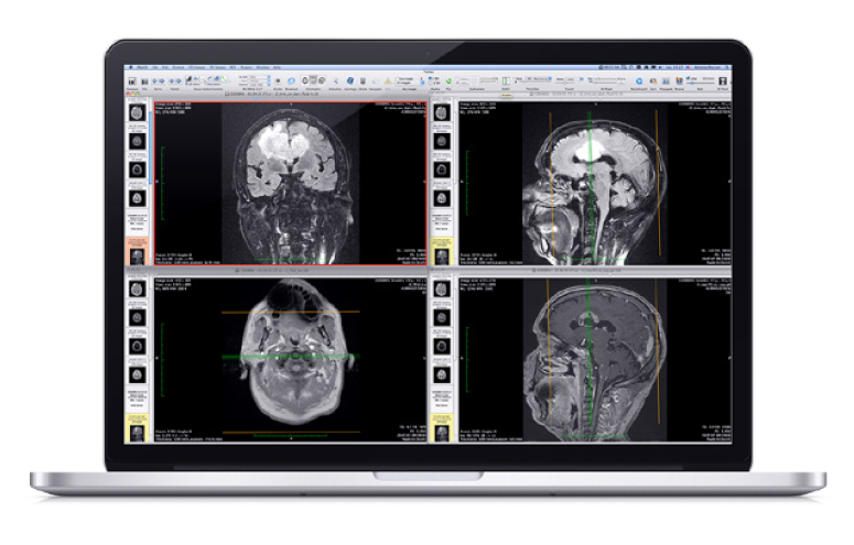
OsiriX MD includes an intuitive interface to display the images. It supports high quality interpolation for best rendering, with Retina screens support.
You can easily add Key Images and Region Of Interests (ROIs) on the images, including lines, polygons, 3D ball, and save them in the database.
You can apply convolutions filter on images, such as bone or lung filters.
OsiriX MD supports 4D images, such as cardiac or perfusion acquisitions and parametric images, such as PET-CT images.
You can define ‘Hanging Protocols’ with multiple screens support.


OsiriX MD offers all the modern post-processing techniques, such as MPR (Multiplanar Reconstruction), 3D Rendering (MIP, Volume Rendering and Surface Rendering).
OsiriX MD supports curved planar reconstruction (3D-MPR) to follow organs such as aorta or bronchi.
You can export 3D reconstructions images as movies, and archive them on your PACS.


OsiriX MD supports DICOM files and also several different types of non-DICOM images, such as LSM files, BioRadPIC, TIFF, ANALYZE, PNG, JPEG, PDF, Quicktime, AVI, MPEG, and more.
OsiriX MD reads and displays all types of DICOM files, produced by medical imaging modalities, including images produced by scanners, MRI, ultrasounds, or standard X-rays.
OsiriX MD can read and display all the DICOM fields associated to the images, such as radiation dose, image position, referring physician, …
OsiriX MD can export DICOM files to CD/DVD or USB sticks, including a stand-alone cross-platform viewer to display the images.
OsiriX MD uses a SQL database to store and index all the images. It can manage several millions of images without problems.


OsiriX MD can receive or send DICOM files, through the DICOM Network protocol.
OsiriX MD can directly print images on DICOM printers.


OsiriX MD is fully optimized for Apple computers, including multi-core processors and graphic board processor support.
OsiriX MD uses asynchronous reading to immediately display the images, even for very large series.

OsiriX MD supports a complete dynamic igins architecture to extend the existing functions.
These nins can directly access the images pixels as 32-bit float for manipulation.
These plugins can create and manage windows, use the entire Cocoa framework, including OpenGL views.
An OsiriX MD plugin is faster than IDL, and easier than ImageJ !
 |
||
|---|---|---|
| OsiriX MD | OsiriX Lite | |
| FDA-Cleared | |
|
| CE IIa Labeled | |
|
| Medical usage | |
“NOT FOR MEDICAL USAGE” |
| User Manual | |
|
| Performances | Up to 80% faster | Standard |
| Email support | |
|
| Pixmeo Website Account | |
|
| Open 500+ images series | |
|
| 2D Images Viewer | |
|
| 3D MPR | |
Demo |
| 3D Curved MPR | |
Demo |
| 3D Rendering | |
Demo |
| Local Database | |
Demo |
| Web Server | |
Demo |
| Web Server user limit | Unlimited | 2 nodes max |
| DICOM Services | |
Demo |
| DICOM Nodes limit | Unlimited | 2 nodes max |
| DICOM Editing | |
|
| Bonjour Protocol | |
Demo |
| CD Creation | |
Demo |
| PET-CT Display | |
Demo |
| 32-bit Pixel Pipeline | |
Demo |
| 11-bit Monitor Support | |
|
| JPEG2000 DICOM | |
Demo |
| Other | |
Demo |
| Test | Results | OsiriX MD | ||||||
|---|---|---|---|---|---|---|---|---|
|
3D Growing Region to segment the colonic lumen Colonic CT 1mm/1mm, 965 images MacPro, 8 cores, 2.8 GHz, 6GB |
|
4.4× faster |
||||||
|
Bone Removing Segmentation in 3D Volume Rendering CTA Lower Limbs 1mm/1mm, 1020 images iMac, 2.8 GHz, 4GB |
|
3.9× faster |
||||||
|
Bone Removing Segmentation in 3D Volume Rendering CTA Lower Limbs 1mm/1mm, 1020 images MacPro, 8 cores, 2.8 GHz, 6GB |
|
4.2× faster |
||||||
|
360 Rotation in 3D Volume Rendering Thoracic CT 1mm/1mm, 760 images iMac, 2.8 GHz, 4GB |
|
1.6× faster |
||||||
|
Loading a large series CTA, multiple series MacPro, 8 cores, 2.8 GHz, 6GB |
|
4.6× more images |
||||||
|
Loading a large series CTA, multiple series iMac, 2.8 GHz, 4GB |
|
2.3× more images |
| Test | OsiriX Lite | OsiriX MD | |
|---|---|---|---|
|
3D Region Growing Segmentation 3D Region Growing to segment the colonic lumen. Colonic CT 1mm/1mm, 965 images. MacPro, 8 cores, 2.8 GHz, 6GB. |
22 sec | 5 sec |
4.4× faster |
|
3D VR Bone Removal Segmentation Bone Removing Segmentation in 3D Volume Rendering. CTA Lower Limbs 1mm/1mm, 1020 images. iMac, 2.8 GHz, 4GB. |
128 sec | 31 sec |
3.9× faster |
|
3D VR Bone Removal Segmentation Bone Removing Segmentation in 3D Volume Rendering. CTA Lower Limbs 1mm/1mm, 1020 images. MacPro, 8 cores, 2.8 GHz, 6GB. |
38 sec | 9 sec |
4.2× faster |
|
3D Volume Rendering 360° Rotation in 3D Volume Rendering. Thoracic CT 1mm/1mm, 760 images. iMac, 2.8 GHz, 4GB. |
138 sec | 85 sec |
1.6× faster |
|
Loading a large series Loading a large series. CTA, multiple series. MacPro, 8 cores, 2.8 GHz, 6GB. |
1’400 img | 6’500 img |
4.6× more images |
|
Loading a large series Loading a large series. CTA, multiple series. iMac, 2.8 GHz, 4GB. |
1’400 img | 3’200 img |
2.3× more images |

For the Brazilian market, an ANVISA version is available. Our Brazilian partner XirisA can provide information and support. Contact XirisA .
Buy OsiriX MD ANVISA (Brazil only)